 2 EP
2 EPEpisodes
Transcript
Episode 1 · Stain, Step, Structure
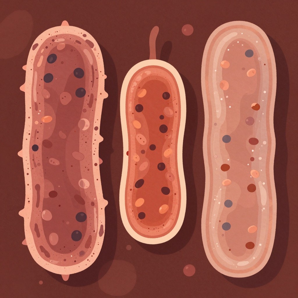
Maya: Purple or pink... and no, this is not a paint-color debate. It’s how a Gram stain gives us a fast clue about a bacterium’s cell envelope. Ezra: Welcome to Purple vs Pink: Gram Basics. Today: stain, step, structure. What are we actually separating here? Maya: Gram-positive from Gram-negative bacteria. The stain sorts them by how their cell walls hold onto color. Ezra: Four steps, in order. Crystal violet first, so everything starts purple. Maya: Then iodine, which helps lock the crystal-violet complex in place. Ezra: Then comes the make-or-break step: alcohol or acetone decolorization. Maya: Exactly. Gram-positive cells have one thick, sturdy peptidoglycan coat... no outer membrane. Think of a big padded purple coat. Ezra: And that thick peptidoglycan traps the violet-iodine complex during decolorization. Gram-positive walls also contain teichoic and lipoteichoic acids. Maya: Gram-negative cells are more like a layered jacket: a thin peptidoglycan layer plus an outer membrane. Alcohol strips the purple complex out, so they become colorless. Ezra: Then safranin comes in as the counterstain. Those decolorized Gram-negative cells turn pink. Maya: Picture a lab tech at 100x oil: purple cocci still visible after decolorization. Their first thought? Thick single-coat wall. Gram-positive. Ezra: That quick distinction matters. It guides lab identification and can shape antibiotic decisions, because the two envelope structures behave differently. Maya: Purple means thick wall, pink means layered jacket. Simple... but seriously useful. Ezra: Next time, we’ll zoom in on what those structures mean beyond the slide. Maya: See you then... and keep staining smart.
Episode 2 · Outer Membranes and Clinical Clues
Maya: Last time, in Stain, Step, Structure, we sketched the bacterial wall. Today, the Gram-negative cell shows up wearing an extra jacket. That’s the whole plot. Ezra: And it is not a fashion detail. Gram-positive bacteria have thick peptidoglycan and stain purple. Gram-negative cells have a thin, about 2 to 8 nanometer, peptidoglycan layer plus an outer lipid membrane containing LPS. Maya: Right. Picture Gram-positive as a big purple sponge. Gram-negative is more like a thin mesh shirt under a raincoat. And, uh, no teichoic acids in that Gram-negative setup. Ezra: Quick stain logic: crystal violet, iodine, then alcohol or acetone. The decolorizer disrupts the Gram-negative outer membrane, and that thin peptidoglycan can’t hold onto the purple complex. Maya: So when safranin arrives, pink or red takes over. Meanwhile, the thick Gram-positive wall hangs onto purple. Tiny color change, huge clue. Ezra: Especially clinically. LPS has endotoxin activity, which helps explain shock-like features in serious Gram-negative infections. Think E. coli, Salmonella, Pseudomonas... different organisms, same important outer-membrane theme. Maya: And that raincoat can be a drug barrier, making Gram-negative bacteria harder to treat with some antibiotics. Ezra: Generally, Gram-positive bacteria are often more susceptible to penicillin-like cell-wall targets. But, hey, don’t prescribe from a stain alone. Check the culture and susceptibility results. Maya: Purple sponge, pink raincoat. There’s your memory hook. Ezra: See you next time.